Our Office
Visit Us Online

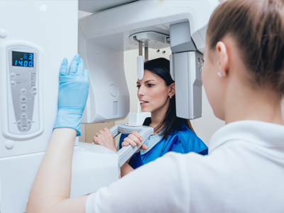
Digital radiography replaces traditional film with electronic sensors that capture X-ray images and transfer them instantly to a computer. For patients, that means faster visits and fewer delays while the clinical team reviews images. The process is streamlined: images are acquired, checked, and stored within moments, allowing your dentist to move quickly from image capture to diagnosis without waiting for chemical processing or film development.
Beyond speed, digital radiography improves the clarity and versatility of images. Clinicians can adjust contrast, zoom in on areas of concern, and apply image-enhancement tools that help reveal fine details. Those capabilities support more confident decision-making during routine exams and when evaluating restorative needs, helping ensure that treatment recommendations are based on the best possible visual information.
At Frisco Smiles Dentistry, we use digital X-ray systems as part of a broader commitment to modern, patient-focused care. The technology supports clear communication: clinicians can share images on-screen with patients, explain findings in real time, and include visuals in treatment planning conversations so patients better understand their oral health.
Digital sensors convert X-ray photons into electronic signals that create a highly detailed image. Compared with film, these sensors capture subtle differences in density and structure, making it easier to detect early-stage problems such as tiny cavities between teeth or the beginning signs of bone loss. Improved image resolution helps clinicians identify issues that might otherwise be missed until they become more serious.
Modern dental software also provides tools to manipulate images without altering the original data. Clinicians can change brightness, enhance contrast, and measure distances on-screen—functions that make assessments more precise. These non-destructive edits preserve the original file while giving practitioners additional views and perspectives for diagnosis and documentation.
Because images are digital, they can be archived in a patient’s electronic record and retrieved instantly during future visits. This historical record allows for side-by-side comparisons over time, making it simpler to track healing, the progression of disease, or the success of previous treatments.
One of the most practical benefits of digital radiography is speed. Instead of waiting for film to be developed, the care team can view an X-ray immediately and begin clinical evaluation. That efficiency shortens appointment times, reduces the need for return visits solely for imaging, and accelerates the path from problem identification to treatment planning.
Instant images also facilitate coordinated care among dental professionals. If a specialist consultation or lab work is needed, digital files can be sent quickly and securely, helping other clinicians review the same high-quality images without delay. This collaborative workflow supports more accurate diagnoses and a smoother patient experience when multiple providers are involved.
For patients, the practical benefit is clearer communication. Clinicians can pull up images during an exam, point to specific areas using on-screen tools, and explain proposed options while the visuals are fresh in everyone’s mind. That transparency helps patients make informed choices about their oral health with a better understanding of the clinical rationale behind recommendations.
Digital radiography reduces radiation exposure compared with traditional film X-rays because modern sensors are more sensitive and require less energy to produce diagnostic images. This lower exposure benefits patients across all age groups, particularly those who need periodic imaging as part of ongoing preventive care or complex treatment monitoring.
Another safety feature is the ability to limit repeat exposures. Since clinicians can immediately verify image quality, mistakes that once required retakes are less common. When a retake is necessary, the team can make small positioning adjustments or change settings on the spot to capture the needed detail without unnecessary repeats.
Digital systems also eliminate the need for chemical processing and physical film, reducing the environmental impact associated with traditional radiography. No developer chemicals, fixers, or film waste are produced, and digital files take up no physical storage space—both practical advantages for a modern dental practice committed to responsible operations.
Finally, secure digital storage supports long-term recordkeeping while enabling controlled access. Image files can be backed up and protected within an electronic health record system, providing durability and privacy protections that paper film archives cannot match.
Digital radiography is designed to work alongside other clinical technologies. Intraoral cameras, digital impressions, and practice management software all exchange information, creating a cohesive digital workflow that improves efficiency and clinical clarity. When systems are integrated, images captured during an exam become part of a complete digital chart used throughout diagnosis, treatment, and follow-up.
This interoperability is especially valuable for restorative and implant planning. Digital radiographs can be combined with other scans and records to guide precise treatment steps, communicate specifications to dental laboratories, and verify outcomes. The result is tighter coordination and improved predictability for procedures that depend on accurate visual and dimensional data.
For patients, integration means more streamlined appointments and clearer explanations. Clinicians can show how X-rays relate to intraoral photos or digital impressions, offering a multi-layered view of oral health that supports shared decision-making. This connected approach helps ensure that clinical recommendations are thorough, well-documented, and easy for patients to follow.
In summary, digital radiography brings faster imaging, clearer diagnostics, enhanced safety, and seamless integration into modern dental care. If you’d like to learn more about how this technology is used in our office or how it can support your oral health, please contact us for additional information.
Digital radiography uses electronic sensors to capture X-ray images that are converted into digital files almost instantly. The sensor detects X-ray photons and converts them to electronic signals, which are processed by computer software to produce a detailed image for clinical review. Because the image appears on-screen within seconds, clinicians can evaluate results without waiting for chemical processing or film development.
This digital workflow makes it easier to enhance and analyze images using tools that adjust contrast, zoom, and measure structures. Those capabilities help clinicians detect subtle changes in tooth and bone density that might be harder to see on film. The result is a faster path from image capture to diagnosis and treatment planning.
Digital sensors generally capture a wider range of detail and subtle density differences than traditional film, which helps clinicians identify early-stage problems such as small interproximal cavities or early bone changes. Image-enhancement tools allow for careful inspection without changing the original file, so clinicians can view the same data from multiple perspectives. Those enhancements support more confident decision-making during routine exams and when evaluating restorative needs.
Another advantage is the ability to compare images side by side over time by retrieving archived files from a patient record. This chronological view makes it easier to track progression or healing after treatment. Together, higher-resolution captures and historical comparisons contribute to more precise monitoring and earlier intervention when needed.
Digital radiography typically requires lower radiation doses than conventional film because modern sensors are more sensitive and capture diagnostic images with less exposure. Clinicians follow established safety protocols, including appropriate shielding and exposure settings that consider a patient’s size, age, and clinical need. Those measures reduce overall exposure while still providing images suitable for accurate diagnosis.
Immediate image review also reduces the need for repeat exposures because clinicians can confirm image quality at the time of capture and make positioning adjustments if necessary. For patients who require periodic imaging, the reduced dose and careful technique together help maintain safety across multiple visits. If you have specific concerns about radiation, your clinician can explain the safeguards used for your care.
Because images are available instantly, appointments tend to be shorter and more efficient, reducing the time patients spend waiting for results. The clinical team can review images on-screen immediately and move directly to discussion or treatment planning without a separate visit for film development. This efficiency streamlines care and minimizes the number of appointments needed for diagnosis and follow-up.
Digital imaging also enhances communication because clinicians can show patients their X-rays on a monitor, point out areas of interest, and explain findings using visual aids. Seeing images alongside intraoral photos or treatment plans helps patients understand recommended care more clearly. The combination of faster results and clearer explanations typically improves patient confidence in clinical decisions.
Digital radiographs are stored as electronic files within a secure patient record system where they can be organized, archived, and retrieved quickly for future comparison. Files are typically backed up regularly to prevent data loss and to maintain a durable clinical history that supports long-term treatment monitoring. Secure storage also eliminates the need for physical film archives and the environmental impact of chemical processing.
Access to digital images is controlled by practice policies and software safeguards that protect patient privacy and meet professional standards. At Frisco Smiles Dentistry, clinicians maintain image confidentiality through secure systems and role-based access to records. If patients have questions about data security or how images can be shared with a specialist, the clinical team can explain the procedures used to protect their information.
Yes. Digital radiographs are commonly used to assess tooth structure, root anatomy, and surrounding bone, which are critical inputs for restorative and implant planning. High-resolution images help clinicians evaluate margins, detect underlying decay, and measure available bone, providing valuable information that guides clinical decision-making and treatment sequencing. Those diagnostic details improve the predictability of restorative outcomes.
When combined with other digital records—such as intraoral scans or cone-beam CT in more complex cases—digital radiographs become part of an integrated dataset that supports precise planning. This interoperability facilitates communication with dental laboratories and specialists and helps verify that restorations and implants meet clinical specifications. The result is tighter coordination and improved predictability for procedures that depend on accurate visual and dimensional data.
Digital radiography is designed to interoperate with intraoral cameras, digital impression systems, and practice management software to create a cohesive digital workflow. Images can be combined with other digital records to produce composite views useful for diagnosis, treatment planning, and patient education. Integration streamlines documentation and reduces the need to manage separate physical records.
For patients this means a more coordinated experience: clinicians can show how X-rays relate to intraoral photos or scanned impressions during the same appointment. That multi-layered view supports shared decision-making and helps ensure that clinical recommendations are well documented and easier to follow. Integrated systems also improve efficiency for clinicians when coordinating care with specialists or laboratories.
Because clinicians can verify image quality immediately, the frequency of repeat exposures due to processing errors or poor film development is markedly reduced with digital systems. Immediate feedback allows small positioning adjustments to be made on the spot if an image does not capture the necessary detail. This capability lowers the incidence of unnecessary retakes and contributes to overall dose reduction.
However, the need for follow-up or additional imaging is determined by clinical indications rather than the imaging medium. Your clinician will recommend imaging at intervals appropriate to your oral health status, treatment progress, and diagnostic needs. In many cases, the improved image quality and instant review offered by digital radiography make monitoring more efficient and informative.
While digital radiographs are excellent for many diagnostic tasks, they have limitations in representing three-dimensional structures on a two-dimensional image. In cases that require detailed three-dimensional assessment—such as complex implant planning, evaluation of certain pathology, or assessment of facial anatomy—a cone-beam CT or other advanced imaging modality may be recommended. Clinicians choose the imaging type that best matches the diagnostic question and clinical risk-benefit profile.
Digital radiography remains a primary tool for routine examinations and many restorative assessments, but clinicians will identify situations where supplemental imaging adds value. If additional imaging is advised, your dental team will explain why it is necessary and how it contributes to a more complete diagnosis or treatment plan. The goal is always to use the least invasive, most informative modality appropriate for your care.
Preparing children or anxious patients begins with clear, age-appropriate explanations and demonstrations of what to expect during the imaging process. Clinicians and staff often use simple language, show the sensor so patients know it is not harmful, and describe the steps of positioning and briefly holding still to obtain a clear image. For many children, a short demonstration or a practice run without radiation helps reduce fear and build trust.
For anxious adults, staff can offer additional time, calming techniques, and positioning adjustments to enhance comfort during the exam. Because digital images are captured quickly, the actual exposure time is brief, which can ease anxiety about prolonged procedures. If special accommodations are needed, the clinical team will work with the patient to make the process as comfortable and efficient as possible.