Our Office
Visit Us Online

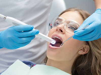
An intraoral camera is a small, pen‑sized imaging device that captures high‑resolution, full‑color pictures from inside the mouth. Designed to reach areas that are difficult to view with the naked eye, the camera transmits real‑time images to a chairside monitor so patients and clinicians can see the same detailed view. This capability transforms a routine exam into a visual conversation, making it easier to identify concerns and explain treatment needs.
Unlike traditional mirrors or written descriptions, intraoral images provide a clear, objective record of tooth surfaces, gum tissue, and restorations. The camera can reveal early signs of decay, hairline fractures, wear patterns, and soft‑tissue changes that benefit from closer monitoring. High‑quality images help clinicians track subtle changes over time and maintain a more accurate clinical history.
Modern intraoral cameras are compact, ergonomically designed, and gentle for patients. They often incorporate LED lighting and macro lenses to produce crisp images without discomfort. Because the device is noninvasive and quick to use, it can be integrated seamlessly into routine checkups, hygiene visits, and focused problem‑solving appointments.
During a typical exam, the clinician positions the camera to capture targeted views of individual teeth, restorations, or areas of concern. The live feed appears immediately on a screen, allowing both patient and clinician to examine the image together. This immediacy speeds up diagnosis and helps ensure that observations are accurate and mutually understood.
Captured images can be saved directly into a patient’s digital record. These files become part of a longitudinal dental file that supports clinical decisions at subsequent visits. When combined with digital radiographs and intraoral scans, camera images create a comprehensive visual archive that guides preventive care and restorative planning.
Because the process is rapid and low‑impact, intraoral imaging is useful across many appointment types: from routine cleanings to focused assessments for sensitivity, bite issues, or new complaints. The practicality of the camera makes it a daily tool rather than a specialty device, improving the consistency and depth of clinical documentation.
One of the primary strengths of intraoral cameras is earlier problem detection. Small lesions and surface irregularities that are easy to miss during a cursory exam often show up clearly on magnified, well‑lit photos. Identifying issues at an earlier stage increases the range of conservative treatment options and can prevent more extensive work down the road.
Intraoral cameras are particularly effective for spotting hairline cracks, marginal gaps around restorations, recurrent decay, and initial enamel breakdown. They also help clinicians assess gum health by documenting inflammation, recession, and plaque accumulation in a way that’s easy to review and compare over time. When combined with periodontal probing and radiographs, camera images complete the diagnostic picture.
Early visual documentation supports evidence‑based treatment planning. By comparing sequential images, clinicians can determine whether a lesion is stable, progressing, or responding to hygiene improvements. That objective comparison reduces uncertainty and supports clinical recommendations rooted in observable change rather than conjecture.
Seeing is often the most persuasive form of communication. When patients view intraoral photographs alongside their dentist, abstract concerns become concrete and easier to understand. This shared perspective improves informed consent, reduces anxiety about the unknown, and empowers patients to participate actively in care decisions.
Clear images help clinicians explain why a recommended procedure is necessary, what alternatives exist, and what outcomes are realistic. Visual evidence can also be used to demonstrate the effectiveness of preventive measures, such as improved brushing technique or targeted flossing, by showing plaque reduction or tissue recovery across visits.
Using this technology fosters trust and transparency. When patients are shown precise images of their oral condition, conversations about priorities and timing become more productive. This collaborative approach supports better adherence to home care instructions and helps patients feel confident that their treatment plan is based on observable findings.
Digital images captured with an intraoral camera integrate smoothly into electronic health records and can be exported as secure image files when collaboration is needed. Specialists, laboratories, and other members of the care team benefit from clear visual references that streamline consultations and laboratory communication. Accurate visuals reduce guesswork and improve the efficiency of interdisciplinary treatment planning.
For restorative procedures, detailed intraoral photographs assist in shade selection, margin assessment, and laboratory communication for crowns, inlays, and other prosthetics. When combined with digital impressions and radiographs, camera images help create a coordinated, predictable workflow that supports high‑quality outcomes.
Privacy and secure storage are important considerations. Images saved to a patient’s chart are handled according to professional standards for recordkeeping and patient confidentiality. Clinicians only share images with other providers or third parties when authorized, ensuring that visual records serve clinical needs without compromising patient privacy.
At the practice level, maintaining a robust visual record supports long‑term continuity of care. New team members can review historical images to understand a patient’s dental history quickly, and patients receive consistent guidance based on documented findings rather than memory alone.
Wrap‑up: Intraoral camera technology brings clarity, efficiency, and collaboration to modern dental care. By making internal views of the mouth accessible to both clinician and patient, it supports earlier detection, clearer education, and stronger treatment planning. At Frisco Smiles Dentistry, we incorporate advanced visualization tools as part of a patient‑centered approach to oral health. Contact us for more information about how intraoral imaging fits into routine exams and your individualized care plan.
An intraoral camera is a compact, pen-sized imaging device that captures high-resolution, full-color photos from inside the mouth. It uses a macro lens and integrated LED lighting to produce clear images of tooth surfaces, restorations, and soft tissues. The live feed appears on a chairside monitor so clinicians and patients can review the same visual information in real time.
The clinician positions the camera to document areas of interest and can save images directly to the patient record for later comparison. Because the device is noninvasive and quick to use, it fits naturally into routine exams and focused problem-solving visits. These still images and short videos create an objective visual record that supports diagnosis and patient education.
During a routine exam the clinician uses the intraoral camera to capture targeted views of individual teeth, margins of restorations, and areas of soft-tissue change. The images are displayed immediately so the clinician can discuss findings with the patient, improving clarity and mutual understanding. Captured photos are stored in the digital chart and can be compared across visits to monitor change over time.
Because imaging is rapid and minimally intrusive, it is often performed alongside visual inspection, periodontal probing, and digital radiographs as part of a comprehensive assessment. This combination gives a fuller picture of oral health and helps prioritize preventive or restorative steps. At Frisco Smiles Dentistry we integrate intraoral imaging into standard documentation to support consistent follow-up and patient communication.
An intraoral camera excels at revealing surface-level issues that are hard to see with the naked eye, such as initial enamel breakdown, hairline cracks, marginal gaps around fillings, and localized plaque accumulation. It can also document soft-tissue changes like inflammation, recession, or unusual lesions that merit closer attention. High-quality images make it easier to spot subtle changes that might otherwise be overlooked during a cursory exam.
While the camera provides excellent surface detail, it is most powerful when used with other diagnostic tools; digital radiographs and intraoral scans reveal issues beneath the surface or between teeth. Sequential photos allow clinicians to determine whether a finding is stable or progressing, which can expand conservative treatment options. This objective visual history supports evidence-based decisions rather than relying on memory or brief observation alone.
Yes. Intraoral cameras are designed to be gentle and ergonomically shaped for patient comfort, with smooth housings and small heads that minimize gagging or discomfort. Imaging is noninvasive and typically takes only a few seconds per view, requiring no anesthesia or special preparation. The device’s LED lighting provides clear illumination without causing heat or pain.
Clinicians follow standard infection-control protocols when handling and positioning the camera to maintain patient safety. Disposable barriers or manufacturer-recommended sterilization methods are used to prevent cross-contamination between patients. These practices make intraoral imaging a low-risk adjunct to routine dental care.
Images captured with an intraoral camera are saved to the patient’s electronic chart as part of the clinical record and are managed according to professional recordkeeping and privacy practices. Digital files can be indexed, timestamped, and associated with specific clinical notes to create an organized visual history. Access to these records is limited to authorized team members to maintain confidentiality.
When images need to be shared with a specialist or laboratory, clinicians export secure image files and obtain patient authorization as appropriate. The practice’s privacy procedures ensure that visual records are used solely for clinical collaboration and treatment planning. This controlled workflow protects patient information while enabling efficient communication among care providers.
Clear intraoral photographs provide objective visual references that streamline communication with specialists, dental laboratories, and other members of the care team. Photos can show shade relationships, margin conditions, and occlusal wear patterns that are useful when fabricating crowns, inlays, or other restorations. Sharing precise images reduces ambiguity and helps external partners understand clinical needs more quickly.
Exportable image files also support remote consultations and case review, allowing team members to evaluate pictures before an in-person visit. This preparation can improve the efficiency of referrals and laboratory workflows by clarifying expectations up front. Ultimately, better visual documentation leads to more predictable restorative outcomes and smoother interdisciplinary coordination.
An intraoral camera is a complementary diagnostic tool rather than a replacement for radiographs or intraoral scans. Cameras excel at documenting surface details and soft-tissue appearance, but they cannot visualize bone structure, root anatomy, or interproximal decay beneath contact points. Radiographs and digital scans remain essential for detecting subsurface pathology and planning many restorative or surgical treatments.
Used together, intraoral photographs, radiographs, and digital impressions provide a comprehensive diagnostic dataset that enhances accuracy and treatment planning. The camera adds contextual surface detail that supports interpretation of radiographic findings and scan data. Combining these modalities leads to more informed clinical decisions than any single tool alone.
The process is straightforward and typically integrated into a routine exam or hygiene visit; the clinician will ask you to sit comfortably and then position the camera briefly to capture the needed views. Each image requires only a few seconds and patients can usually breathe and swallow normally throughout the procedure. Most people experience no discomfort, and children often tolerate the imaging well with minimal explanation.
After images are taken, the clinician will review them with you on a monitor and explain any findings using the photos as a visual aid. This discussion helps you understand recommended next steps and lets you ask questions with the image visible for reference. Saved photos become part of your chart so you and your clinician can track progress at future visits.
Photographic documentation makes oral hygiene issues tangible by showing plaque accumulation, gingival inflammation, and localized wear that can improve with targeted home care. When patients see images that illustrate areas missed during brushing or flossing, clinicians can demonstrate technique adjustments more effectively. Visual feedback often motivates patients to adopt better habits because progress can be tracked photographically over time.
By comparing sequential images, clinicians can evaluate the effectiveness of preventive measures and reinforce successful behavior changes. This objective monitoring helps guide personalized hygiene instructions and periodic professional interventions. As a result, intraoral imaging contributes to earlier correction of risk factors and stronger long-term oral health outcomes.
Maintaining reliable intraoral imaging requires regular equipment checks, manufacturer-recommended calibration, and routine cleaning or barrier use between patients. Staff training is also essential so team members capture consistent, diagnostically useful images and integrate photos properly into the digital record. Technology updates and software patches are applied as needed to ensure compatibility with practice management systems and imaging workflows.
Investing in ongoing maintenance and training helps the practice keep imaging accuracy high and support efficient care coordination with specialists and laboratories. When new camera features or improved imaging software become available, clinicians evaluate them for clinical benefit before adoption. Frisco Smiles Dentistry prioritizes these practices to keep visualization tools current and reliable for patient care.