Our Office
Visit Us Online

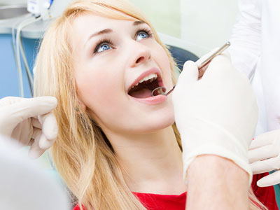
At Frisco Smiles Dentistry, we take a preventive-first approach because the best dentistry is often the care you never wish you'd needed. Regular oral exams give our team the information we need to protect your teeth, gums, and overall oral function before small issues become larger problems. During an exam, we combine a careful clinical inspection with targeted diagnostics, clear communication, and a personalized plan so each patient understands the condition of their mouth and the steps to preserve it.
Your first comprehensive oral exam with our team establishes a baseline for future care and helps us learn about your health history, habits, and goals. We begin by reviewing medical and dental histories and asking about current concerns—sensitivity, pain, changes in bite, or aesthetic worries. This conversation guides the physical exam and any additional tests we recommend so you leave with clarity about your oral health.
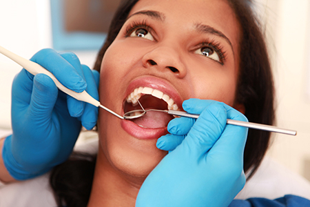
The clinical portion of the exam inspects every accessible surface of the mouth: teeth, gum tissue, tongue, cheeks, and the soft tissues of the throat and neck. We check for signs of decay, inflammation, abnormal lesions, and wear patterns that could indicate grinding or clenching. We’ll assess your bite and observe the temporomandibular joint (TMJ) for limited movement, clicking, or discomfort that can affect daily function.
When indicated, diagnostic images help reveal what can’t be seen with the naked eye. Digital x-rays or other imaging can show early cavities between teeth, root and bone health, and the presence of impacted or developing teeth. After the exam, we’ll review findings with you, explain recommended next steps if any, and outline options so you can make informed decisions about your care.
The mouth is a window to general health. During routine exams, our team watches for oral signs that can be related to systemic conditions—ranging from nutritional deficiencies and hormonal changes to chronic inflammatory diseases. Recognizing these connections helps us coordinate care and, when appropriate, recommend that patients consult with their medical providers for evaluation of broader health concerns.
Common symptoms that may have causes beyond the mouth include persistent dry mouth, unusual bleeding, unexplained sores or lumps, and sudden changes in taste or smell. While these findings are not diagnoses in themselves, they often prompt additional assessment or referrals when they suggest an underlying issue. Early detection in the oral cavity can sometimes lead to earlier attention to health problems elsewhere in the body.
Mounting research highlights links between oral disease and conditions such as cardiovascular disease, diabetes, respiratory illnesses, and adverse pregnancy outcomes. During an oral exam we not only screen for dental disease, but also document signs that could indicate medical conditions, helping you and your healthcare team take a more connected approach to overall wellness.
Daily brushing and flossing form the foundation of good oral health, but professional cleanings remove hardened tartar and bacterial buildup in places that home care can’t always reach. Our hygienists use specialized instruments and techniques to clean below the gumline when necessary, polishing teeth and advising on targeted improvements to your home routine. This partnership between in-office care and consistent home habits is the most effective way to prevent cavities and gum disease.
Regular cleanings also allow us to monitor subtle changes over time. Gum pockets, areas of enamel erosion, or persistent staining can indicate behaviors or health factors that need attention. By catching these trends early, we can often intervene with conservative measures rather than progressing to more involved treatment.
We generally encourage patients to maintain regular checkups and cleanings at intervals recommended for their individual risk profile. For many people that means twice-yearly visits, while others may need more frequent monitoring. During your appointment, our hygienist will demonstrate proper brushing and interdental cleaning techniques and tailor preventive advice to your lifestyle and needs.
Children benefit particularly from a consistent preventive program. Early exams and professional cleanings help establish good habits, protect developing teeth, and give parents guidance on nutrition, fluoride use, and injury prevention to support long-term oral health.
Imaging is an essential part of many oral exams because it reveals internal structures that a visual inspection cannot. Digital radiographs show the extent of decay, the health of tooth roots and surrounding bone, and the position of unerupted teeth. These images guide accurate diagnoses and ensure that treatment plans address the full picture of a patient’s oral health.
Advances in digital radiography have made imaging faster, more convenient, and more precise. Digital sensors produce high-resolution images instantly, allow for image enhancement to clarify findings, and minimize radiation exposure through more efficient technology. Images can also be stored in a patient’s electronic file for comparison over time, which supports continuity of care and long-term monitoring.
Imaging is selected based on clinical need and patient history. For example, small intraoral films detect early interproximal decay; panoramic images offer a broad view of jaws and erupting teeth; and three-dimensional scans provide detailed anatomic information for complex cases. We always explain the purpose of any image we recommend and how it contributes to your diagnosis and treatment planning.
The specific type of radiograph chosen depends on your symptoms, medical history, and the clinical questions we need to answer. Each modality provides unique information that helps us detect disease early, plan restorations, and evaluate structures that are not visible during a routine exam.
Periapical x-ray - Focuses on an individual tooth from crown to root and surrounding bone, useful for detecting root issues and localized infections.
Bitewing x-ray - Captures the upper and lower crowns in a single view to detect interproximal decay and monitor existing restorations.
Full mouth series - A comprehensive set of periapical and bitewing images that provides a detailed baseline of the entire dentition.
Panoramic film (panorex) - Offers a broad overview of the teeth, jaws, and adjacent structures; helpful for evaluating development, impacted teeth, and jaw pathology.
Cephalometric film - Provides a profile view of facial bones and is commonly used for orthodontic assessment and growth analysis.
For advanced diagnostic needs—surgical planning, implant placement, or evaluation of complex anatomy—cone-beam computed tomography (CBCT) produces three-dimensional images of the teeth, jaws, and surrounding tissues. CBCT offers a detailed spatial understanding of anatomy that improves surgical precision and treatment predictability when such information is clinically indicated.
Our goal during every oral exam is to use the least invasive diagnostic tools necessary to obtain the information needed for confident decision-making. When imaging is recommended, we’ll explain why it’s helpful, what it will show, and how it influences your care plan.
In summary, a thorough oral exam combines a careful clinical review, targeted diagnostics, and personalized education to maintain and improve oral health. If you have questions about what to expect during an exam or would like more information about any of the screening tools we use, please contact us for more information. We look forward to helping you protect your smile for years to come at Frisco Smiles Dentistry.
An oral exam is a clinical evaluation of the teeth, gums, soft tissues and supporting structures that detects early signs of disease and dysfunction. Exam components typically include a visual inspection, tactile assessment, bite evaluation and a review of medical and dental history. These visits focus on prevention and give the dental team the information needed to recommend timely, conservative care.
Oral exams are important because many dental problems are easier and less invasive to treat when found early. They also provide opportunities to identify signs that may relate to overall health, such as nutritional deficiencies, infections or systemic inflammatory conditions. Regular screening supports long-term oral function, comfort and appearance.
During your initial comprehensive oral exam the dental team establishes a baseline and gathers a full medical and dental history. At the office of Frisco Smiles Dentistry we ask about current symptoms, medications and habits such as grinding or tobacco use to tailor the exam. This intake helps guide any additional tests and ensures your care plan matches your goals and risk factors.
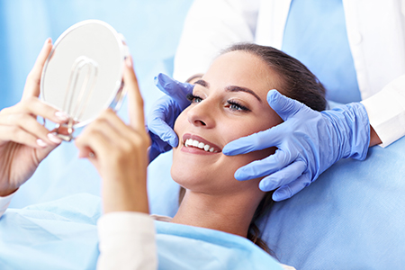
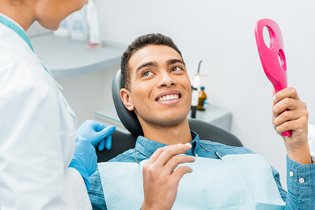
The physical portion of the exam inspects all accessible intraoral and extraoral tissues, including teeth, gums, tongue, cheeks, lips and the temporomandibular joint. Clinicians check for decay, inflammation, abnormal lesions, wear patterns and signs of infection or developmental concerns. Findings are reviewed with you at the appointment so you understand recommended next steps and preventive strategies.
The frequency of oral exams varies by individual risk but many adults benefit from twice-yearly visits for routine screening and maintenance. Patients with active gum disease, a history of decay, certain medical conditions or ongoing dental treatment may need more frequent monitoring. Your dentist will recommend an interval based on your oral status, overall health and lifestyle factors.
Regular scheduling allows the team to track subtle changes over time, compare diagnostic images and intervene before problems progress. More frequent exams can prevent small issues from becoming complex procedures and help manage chronic oral conditions effectively. Children, patients undergoing orthodontic treatment and those with systemic conditions often require tailored intervals.
Oral exams are a primary opportunity to screen for oral cancer and other serious conditions through visual inspection and palpation of soft tissues. Clinicians look for suspicious sores, white or red patches, persistent lumps and asymmetry in the mouth and neck. When findings are unclear the team documents size, location and duration and recommends timely biopsy or medical referral when indicated.
Because the mouth can reflect systemic disease, exam findings such as unusual bleeding, persistent dry mouth or changes in taste often prompt broader health discussions. Coordinating with a patient’s medical providers ensures that suspected systemic issues receive appropriate evaluation and treatment. Early detection through regular exams increases the chances of successful treatment when serious disease is present.
Diagnostic tools commonly used during oral exams include digital radiographs, intraoral cameras and, when needed, three-dimensional imaging such as cone-beam computed tomography. Digital x-rays reveal interproximal decay, root and bone conditions that are not visible on inspection alone. Intraoral cameras allow the clinician to magnify and document lesions or areas of concern for patient education and recordkeeping.
CBCT provides detailed spatial information for complex cases such as implant planning or assessment of anatomical relationships. Selection of imaging and adjunctive tools is always based on clinical need, history and the specific diagnostic question. The team will explain why an image is recommended and how it contributes to accurate diagnosis and treatment planning.
Radiographs are recommended when they will provide information that changes diagnosis or influences treatment decisions, such as detecting early cavities or evaluating root and bone health. Common films include periapical images for individual tooth roots and surrounding bone and bitewing images for interproximal decay. A full mouth series offers a comprehensive baseline when a more detailed assessment is needed, while panoramic films provide an overview of the jaws and developing teeth.
Advanced three-dimensional imaging is reserved for surgical planning, implant placement and complex anatomy assessment. Your dentist will consider your symptoms, clinical findings and prior imaging when recommending radiographs to minimize exposure while maximizing diagnostic value. Digital systems reduce radiation dose and allow instant image enhancement and storage for comparison over time.
Professional cleanings complement oral exams by removing hardened tartar and bacterial buildup from places brushing and flossing can miss. Hygienists use specialized instruments to clean below the gumline when necessary, reducing inflammation and supporting gum health. Polishing and targeted hygiene instruction at these visits also improve aesthetics and patient comfort.
Regular cleanings give the team an opportunity to monitor pocket depths, enamel erosion and staining trends that may indicate changing risk. Based on these observations, clinicians can recommend preventive measures such as fluoride, sealants or behavior modifications to slow disease progression. This collaborative approach between in-office care and consistent home routines is the most effective way to prevent cavities and periodontal disease.
Oral exams for children emphasize growth, development and preventive education while addressing unique pediatric risks such as early decay and injury. Clinicians assess tooth eruption patterns, bite development and habits like thumb sucking or pacifier use that can affect alignment. Early exams also allow parents to receive guidance on nutrition, fluoride use and injury prevention to protect developing teeth.
Children often benefit from more frequent monitoring during periods of rapid dental development or when preventive treatments such as sealants are indicated. Pediatric exams are tailored to be positive and age-appropriate, building habits that support a lifetime of oral health. When orthodontic concerns are present, exams include assessment for timely referral to an orthodontist.
Before your oral exam it is helpful to share a complete medical history, current medications, allergies and any recent hospitalizations or chronic conditions. Informing the team about symptoms such as sensitivity, bleeding, swelling, facial pain or changes in taste helps focus the evaluation. Frisco Smiles Dentistry also asks about habits like tobacco use, night grinding and diet because these factors influence risk and treatment planning.
Tell the dental team about pregnancy, autoimmune conditions or recent changes in medication, as these can affect oral findings and treatment choices. If you have recent dental images or notes from another provider, bringing them to your appointment can speed diagnosis and avoid duplicate imaging. Clear communication ensures your exam is thorough and the recommended plan fits your overall health needs.
After an oral exam recommended follow-up steps may include a professional cleaning, targeted restorative work, periodontal therapy or monitoring with periodic rechecks. Some patients require diagnostic testing or referral to a specialist for biopsy, medical evaluation or advanced treatment. The dentist will prioritize conservative measures when possible and outline options so you can make informed decisions about care.
Follow-up plans include specific timelines and goals to help measure progress and maintain oral health. Education about home care techniques and scheduling of preventive visits are common outcomes of an exam and support long-term stability. Regular follow-through on the agreed plan is the most reliable way to protect oral function, comfort and appearance over time.